Distefano Research Group

Protein Prenylation - The Anchor of Life

Protein prenylation is a post-translational modification that consists of the attachment of 15 or 20 carbon isoprenoids to specific cysteine residues positioned near the C-termini of proteins. In a eukaryotic cell, there are several hundred prenylated proteins including most members of the Ras superfamily and heterotrimeric G-proteins; the prenyl group serves to anchor these proteins in the membrane so that they are positioned to interact with cell surface receptors either directly or via adaptor proteins. This means that essentially all signaling processes in eukaryotic cells require the participation of prenylated proteins for everything ranging from the regulation of cell division to stem cell differentiation and development. Beyond biological significance, the critical role of prenylated proteins also makes them important targets for the design of new therapeutic agents for a variety of diseases.


Research in the Distefano Group
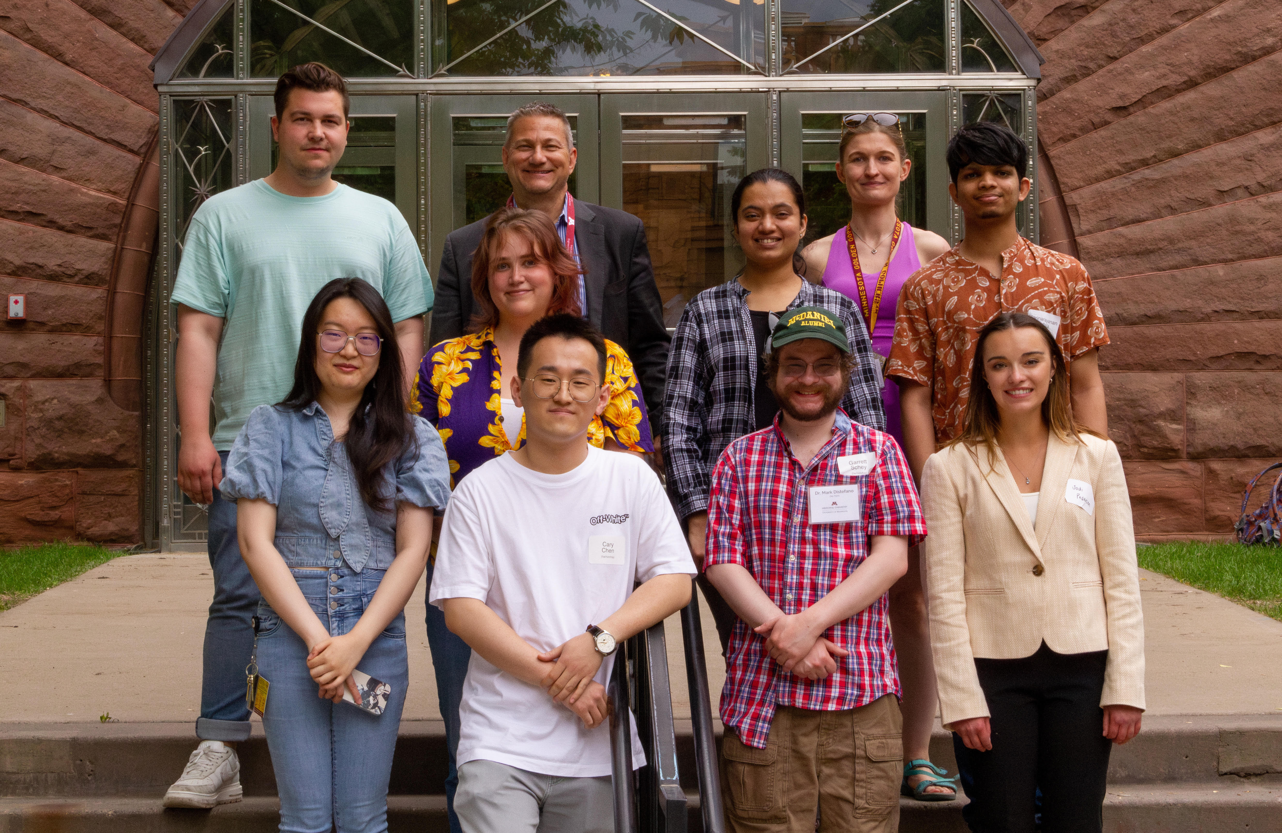
GP230616

Works in the Distefano Group on protein prenylation is focused in two areas: Chemical Biology and Biotechnology Applications. In pursuit of those studies, members of the group perform a variety of different types of experiments including chemical synthesis, biochemistry, proteomics, cell culture and animal-based work.  The goal of these works is to gain insight into protein prenylation that can be used to advance biology and develop new therapeutic approaches for a broad range of diseases including cancer, Alzheimer’s disease and infectious disease.
Research Highlights

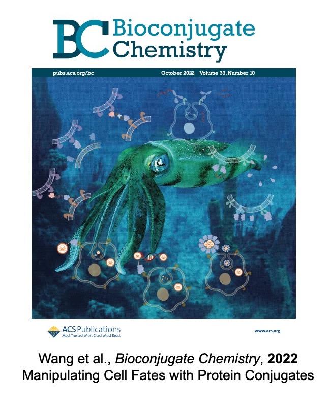
R.H-wang
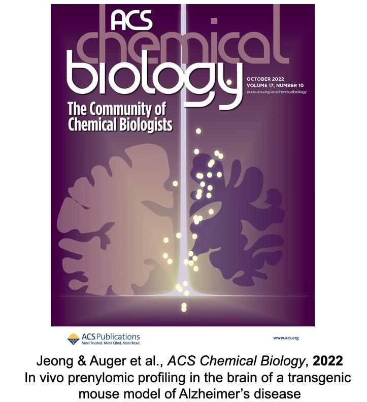
R.H-J-A

MDD Group News

Red-shifted backbone N–H photocaging agents

In a recent article published in Organic and Biomolecular Chemistry, graduate student Taysir Bader, in the research group of Professor Mark Distefano, in collaboration with graduate student Alicia E. Mangubat-Medina, and undergraduates Hallie O. Trial, Reyner D. Vargas, and Mekedlawit T. Setegne in the research group of Dr. Zachary Ball at Rice University, described the synthesis and characterization of two new nitrodibenzofuran (NDBF) based boronic acid reagents for peptide backbone N–H caging and subsequent photocleavage of the resulting C(sp2)-X bond.

The boronic acid reagents were synthesized using an elegant strategy that started with 4-fluoro-2-nitrobenzaldehyde, which was used to construct the dibenzofuran moiety via a C–H activation/cyclization and late-stage introduction of a propargyl group, followed by hydroboration. This strategy was used to produce two boronic acid analogues; one containing the parent NDBF, and the other with a dimethylamino-substituted analogue with a red-shifted absorbance profile that gives further access to two-photon mediated cleavage. With the boronic acids in hand, they were used to protect the backbones of three different peptides: pep1, a collagen-type sequence that exhibits triple-helix folding behavior known to be disrupted by backbone N– H alteration (seq: Ac-(POG)3POGHOG(POG)3-NH2); pep2, a hormone releasing peptide (LHRH) that contains a pyroglutamate–histidine motif (seq: pE– HWSYGLRPG-NH2); and pep3, a manufactured hormone (leuprolide) used to treat several types of cancer (seq: pE-HWSY-DLeu-LRP-NHEt). HPLC and MALDI were used to show clean Cu(NO3)2 mediated caging of these peptides in NMM buffer, and subsequent uncaging upon irradiation with a blue LED light in isoamylamine buffer. Analysis of the uncaging kinetics using pep3 showed that NDBF uncaging (t90% = 37 s, Φ·σ = 4.7 × 106 cm2 mol−1) was 7-fold faster than previous generation nitroveratrole protecting group (t90% = 257 s, Φ·σ = 6.7 × 105 cm2 mol−1), and 23-fold faster than the dimethylamino analogue (t90% = 907 s, Φ·σ = 1.9 × 105 cm2 mol−1), indicating that it disfavors one photon uncaging. Two photon uncaging at 800 nmon the other hand was noticeably faster for the dimethylamino analogue compared to the parent NDBF, with a cross section on the order of 0.13 GM.

Specific RNA splicing inhibitors lead to cancer reduction

In reseach recently published in the Proc. Nat. Acad. Sci. U.S.A., graduate student Kiall Suazo in the Distefano Lab contributed to a collaborative project led by Professor Carol Williams in Medical College of Wisconsin in altering SmgGDS ratios using splice-switching strategy. SmgGDS has two isoforms (SmgGDS-607 and SmgGDS-558) that work together in prenylating and subsequent trafficking of a set of small GTPases. A high 607:558 ratio is implicated in cancer by supporting malignant phenotypes and tumor development. In an effort to reduce the ratio of SmgGDS-607 and SmgGDS-558 in a lung cancer model cell line, a splice-switch oligo nucleotide (SSO) was designed, which works by decreasing the amount of 607 and increasing 558.

Using a chemical proteomics approach developed in the Distefano lab, it was found that oncogenic small GTPases exhibited reduced prenylation upon SSO treatment, indicating that alteration of the SmgGDS ratio inhibits the prenylation of this subset of prenylated proteins. This observed reduce prenylation in cancer cells promotes ER stress, resulting suppressed tumor growth or induced cell death. Furthermore, the results from the proteomics study may have uncovered a novel aspect of SmgGDS intracellular function, linking the 607:558 ratio to the direct regulation and control of prenylation for other types of prenylation substrates. Indeed, this study exemplifies the power of chemical proteomic strategies to uncover aberrant levels of post-translationally modified proteins under perturbed conditions.

Former Postdoc Matt Hammers Starts Faculty Position at UW LaCrosse

Former Distefano group postdoc Matt Hammers started his independent career as a Professor at University of Wisconsin La Crosse this fall.  In the Distefano group, Matt worked on the development of new photoremovable protecting groups for cysteine and their use for biological experiments including the uncaging of peptides and drugs inside cells.